Discovering such new structures from nature, is an essential part of the very first steps of drug discovery and the more diverse the compounds, the better chances we have - only 1 out of 100,000 to 1,000,000 compounds tested will make it into a clinically approved drug, but when it works, it works really well. For example, years ago a sample of soil was collected from Easter Island (Rapa Nui). After culturing the bacteria scientists discovered rapamycin, also known as aka sirolimus, which is today used during organ transplants to prevent the donor organs from being rejected.
But the biggest problem in the field of marine biodiscovery is determining the structure of unknown molecules that might have medical potential - and the more complex they are the longer it takes, sometimes years, thus delaying potential cures. So anything that can speed up this process is an accomplishment all by itself.
Normally we can use high-resolution mass spectrometry and nuclear magnetic resonance techniques to determine the compounds, but in the case of a particular compound from the Mariana Trench that we studied last year we had difficultly resolving its exact molecular structure.
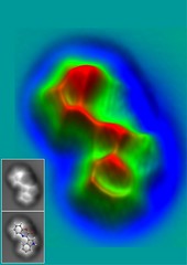
Left with four potential structures, we read in the local newspaper about a recent breakthrough at IBM's lab in Zurich where they imaged for the first time an organic molecule, pentacene, using atomic force microscopy (AFM). This technique, had up to then only been used to image molecules as large (or small) as proteins or peptides. A small side note, the AFM is the offspring of the scanning tunneling microscope, which was given the Nobel Prize of Physics in 1986 - it was also invented at the IBM Zurich lab. After doing some quick searching online and watching this YouTube video I contacted Dr. Leo Gross at IBM and explained to him our problem.
A few months later they had the results -- cephalandole A, which is actually known and originally isolated from a Taiwanese orchid -- interesting enough, it's structure that was published originally had later been revised by total synthesis.